D. Dingli, M-Y Jung, C. P. Offord, et al. Cancer Research, August 2018
Data from high-resolution imaging suggest that methods that enable the quantitation of in vivo anisotropies are required for continuing development of successful oncolytic virotherapies.
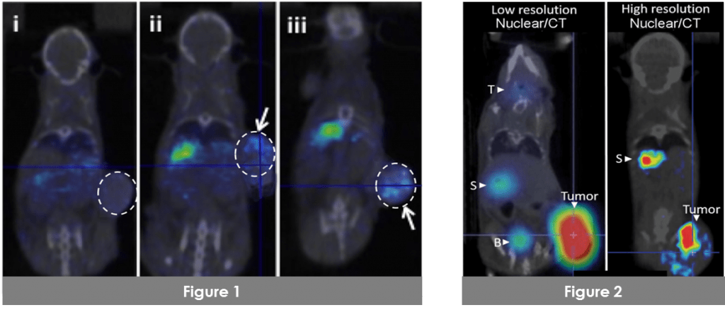
Fig.1: High-resolution U-SPECT/CT imaging of intratumoral isotope uptake in 5mm tumor. In (i) a control tumor has no isotope activity, whereas two representative tumors (ii and iii) infected with MV expressing NIS concentrate the isotope above background. Click here for the full article.
Fig. 2: In another article of Mayo Clinic (A. Miller & S. J. Russell, Expert Opin. Biol. Ther.16(1):15-32) demonstrate that high-resolution nuclear imaging is required in order to identify intratumoral infected oncolytic viral centers required to optimize oncologic treatment. Click here for the full article.
Summary of VECTor6 advantages for whole-body tumor micro-environment imaging:
- The only system that can image all available radiotracers for NIS-mediated imaging at high resolution: 123I, 124I, 125I, 131I, 18F-tetrafluoroborate, 99mTc-pertechnetate, and 188Re.
- With a single acquisition, it is possible to directly compare the spatiotemporal resolution of PET and SPECT tracers such as 124I/18F, 123I/99mTc, 123I/124I, and 99mTc/18F. Knowledge of both spatial distribution and kinetics of infection will allow for better understanding of oncolytic virotherapies and help to inform clinical practice and dose determination.
- Simultaneous imaging of PET and SPECT tracers with both spatial and temporal co-registration makes the translation from preclinical to clinical tracers a one-to-one process.
- The system enables radiovirotherapy with 131I and 188Re at high sub-mm resolution.
