A Pioneering Mind in Nanomedicine and Theranostics
 Prof. Dr. Dr. Twan Lammers is Head of the Department Nanomedicine and Theranostics at the Institute for Experimental Molecular Imaging (ExMI) at the University Hospital Aachen and the Helmholtz Institute for Biomedical Engineering at RWTH Aachen University, Germany.
Prof. Lammers is an expert in drug targeting, image-guided drug delivery, tumor-targeted combination therapies, and molecular imaging. In 2020, he received the International Award of the Belgian Society for Pharmaceutical Sciences. He has been ranked as one of the most highly cited researchers in Biomedical Technology, Nanomedicine and Drug Delivery Systems by ExpertScape. Since 2019, he has also been included in the list of the World’s Most Influential Scientific Minds by Clarivate Analytics. Defining the future of the field, he recently published a perspective on “Smart cancer nanomedicine” [1] as well as a chapter on “Patient stratification to improve nanomedicine performance and clinical translation” in the 2020 Roadmap on Nanomedicine [2].
Thus far, cancer nanomedicine formulations have largely failed to improve therapeutic outcomes in the clinic in terms of patient survival. What is the primary reason for this?
Since 2000, many thousands of papers have been published on cancer nanomedicines, and many big promises have been made. Conversely, only seven cancer nanomedicine products have developed in the past two decades. I do not think that seven is a bad number, especially not for the young field that nanomedicine is, but the number does not live up to the high hopes and big expectations that have been created. In part, this disbalance can be explained by the lacking availability of protocols for patient stratification in cancer nanomedicine. These are needed because of the high inter- and intra-individual heterogeneity that exists in the EPR (i.e. Enhanced Permeability and Retention) effect, on which nanomedicine tumor accumulation is based, and which is often mistakenly assumed to be omnipresent in all tumor types. However, tumors tend to be very heterogeneous by nature and an increasing number of studies has shown that the EPR effect within tumors is also highly heterogeneous. This heterogeneity results from variability in multiple EPR-contributing parameters, such as vessel density, perfusion and permeability, tumor stroma composition, interstitial fluid pressure, and lymphatic vessel density and functionality. To move the field forward, we have to improve our understanding of the EPR effect, of its tumor type-specific pathophysiology, of nanomedicine behavior in the body, and of nanomedicine interactions with the heterogeneous tumor microenvironment. In addition, we have to keep the big picture in mind and consider specific challenges associated with clinical translation and nanomedicinal drug development [3,4].
As a pioneer in the field of nanomedicine and the EPR effect, are there any lessons you would like to share on improving the clinical translation of cancer nanomedicines?
At the preclinical level, integrating imaging in nanomedicine and drug delivery research has enabled researchers the non-invasive and quantitative assessment of nanocarrier biodistribution, target site accumulation, and (triggered) drug release [5]. Clinically, we will have to move toward image-guided and personalized nanomedicine based on the selection of patients on the basis of noninvasive imaging information. Ideally, not only accumulation in primary tumors should be considered, but also localization in metastases. In addition, besides nanomedicine accumulation, also their distribution in cancerous lesions is important and should be captured. Depending on the accumulation and distribution patterns of nanomedicines in tumors and metastases — which as mentioned above tend to vary quite substantially — optimized treatment protocols can be conceived for individual patients.
As one of the most cited researchers in the field of nanomedicine and drug delivery systems, can you elaborate on the imaging techniques used in your laboratory?
To visualize and quantify the circulation behavior, biodistribution, and target site accumulation of cancer nanomedicines, imaging agents are being entrapped within or conjugated to nanoparticles, allowing them to be tracked via non-invasive imaging techniques such as Optical Imaging (OI) and radionuclide imaging.
In terms of Optical Imaging, we use MILabs’ hybrid CT-FLT (Computed Tomography-guided Fluorescence Tomography). This enables CT-based assessment of mouse and organ anatomy together with a near-infrared fluorophore (NIRF)-based optical tomography. To permit non-invasive optical quantification, Dr. Felix Gremse at our ExMI institute has developed scattering and absorption reconstruction protocols, in order to correct for the shape of mice and for the different optical properties of different organs, e.g. the strong absorption of light in highly perfused organs, such as the heart, and the liver [6]. This technology has proven to be very useful in preclinical research, to more meaningfully and more quantitatively assess nanomedicine biodistribution and target site accumulation.
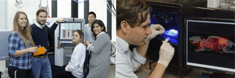
Prof. Lammers and his team extensively use the MILabs’ CT-FLT device, complemented by advanced microscopy
By employing multimodal and multiscale imaging techniques in a complimentary manner, we are able to systematically study the vascular network and the stromal composition in tumors and metastases; we can visualize and quantify nanomedicine-mediated drug targeting to tumors and metastases; and we can non-invasively evaluate macrophage- and vasculature-modulation therapies to enhance tumor-targeted drug delivery and anticancer therapy. In this context, high-resolution micro-CT is oftentimes used to assess the relative blood volume in tumors, as well as vessel size, vessel distribution and vessel branching. In parallel, CT-FLT-based methods allow us to assess the pharmacokinetics, biodistribution and target site accumulation of various different types drug delivery systems, including polymers, micelles and liposomes [7,8,9,10]. Using these techniques together with advanced microscopy as tools to assess the efficacy of macrophage/vasculature-modulation therapies, we furthermore demonstrated that genetic, pharmacological and physical priming of tumor blood vessels and the microenvironment can help to improve tumor-targeted drug delivery [11,12].
Can you comment on your work to enhance the EPR effect?
Let me first clarify that I think it is important to better understand the basics of the EPR effect. We might have been underappreciating the importance and contribution of transcytosis with regard to nanoparticle access to tumors. Analogously, we might have been overrating the importance of absent lymphatics in nanoparticle retention as compared to capture by tumor-associated macrophages. Vascular leakiness (and/or transcytosis) may not be sufficient in certain tumors to ensure good therapeutic efficacy. Unlike preclinical subcutaneous tumor models, which often have homogenous and high levels of vessel leakiness caused by rapid tumor development, human tumors are known to have much more mixed levels of “EPR” and target site accumulation, in part sometimes also caused by poor tumor blood vessel perfusion. Therefore, protocols to enhance tumor perfusion, permeability and penetration appear to be essential to ensure clinical success. Over the years, we and others have been studying multiple clinically relevant pharmacological and physical co-treatments to enhance nanomedicine accumulation. Examples of pharmacological priming are vessel permeabilization, normalization, disruption and promotion, and examples physical priming are hyperthermia, radiotherapy, sonoporation and photodynamic therapy.
Can you elaborate on your further expectations & future directions?
Like with CT-FLT optical imaging to monitor biodistribution and tumor targeting at the preclinical level, we believe that imaging is also very important in the clinical arena, to promote translation and personalize nanotherapies. We are in parallel looking at biopsy biomarkers to predict nanomedicine accumulation and efficacy, as those might be more practicable and more cost-efficient as imaging biomarkers. That said, in patients with metastases — which will be the majority in early-stage clinical trials — imaging seems to be the only available means to promote translation and enable stratification, especially in case of passively targeted nanomedicines. Besides imaging and patient stratification, another important future direction in this area of research is the combination of nanomedicine with immunotherapy. Prominent progress has already been reported in multiple preclinical models and first findings on the efficacy of nano-immunotherapy in patients are eagerly awaited.
References:
[1] Van der Meel, Lammers et al, Nature Nanotechnology 2019
[2] Decuzzi, Lammers et al, Nanotechnology 2020
[3] Lammers, Macro-nanomedicine, J Control Release 2019
[4] Metselaar, Lammers, Drug Deliv Transl Res 2020
[5] Dasgupta, Biancacci, Lammers, Theranostics 2020
[6] Gremse, Theek, Lammers et al, Theranostics 2014
[7] Kunjachan, Lammers et al, Nano Lett. 2014
[8] Tsvetkova, Lammers et al, Nano Lett. 2017
[9] Biancacci, Hu, Lammers et al, J Control Release 2020
[10] May, Golombek, Lammers et al, Theranostics 2020
[11] Ojha, Lammers et al, Adv Drug Deliv Rev 2017
[12] Theek, Baues, Lammers et al, J Control Rel 2018
