PRESS RELEASE
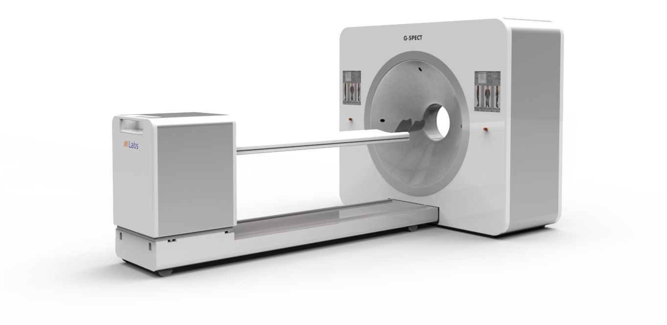
UTRECHT, June 3, 2015 – G-SPECT, a new Single Photon Emission Computed Tomography (SPECT) system developed by MILabs, enables ultra-fast and highly detailed scanning of biological processes, relevant for diagnosis and research in the human body. The unprecedented high SPECT resolution of better than three millimeters is expected to enable physicians to arrive at a more accurate diagnosis. In addition, the G-SPECT will enable the quantification of a great numer of fast dynamic processes in e.g. the brain for the first time.
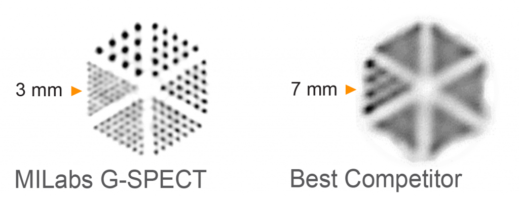
With the G-SPECT from MILabs the scanning speed in the brain improves to about half a minute and in some cases even a fraction of a second. Therefore fast processes can now be visualized for the first time. Also image blurring is minimized (3mm). In comparison, the resolution of existing SPECT scanners is usually 8 to 10 millimeters. Moreover, traditional SPECT machines are equipped with heavy, rotating detectors, one reason why it takes such a long time for a 3D image to be acquired: usually 10 to 30 minutes. G-SPECT works without any rotation of the detectors which will positively impact the wear and tear of the equipment and thus increases reliability.
 |
 |
“Normally SPECT scanners provide a picture and that’s it. With the G-SPECT we put 3D images very quickly one after another, resulting in a 4D movie of exceptional image quality” explains CEO prof. dr. Freek Beekman, also a researcher at the Technical University of Delft, The Netherlands, where a part of the basic research on G-SPECT was conducted. “We expect that doctors can pinpoint disease much earlier and with much greater certainty find abnormalities in the case of epilepsy, Parkinson’s and Alzheimer’s disease, and also differentiate more precisely between patients since tracer uptake in tiny brain structures becomes visible. This can for example be essential to quantify changes in local blood flow, receptor binding and metabolism.” With the G-SPECT MILabs builds on the previously developed high resolution preclinical scanning technology of small animals.
Prof. Fred Verzijlbergen,Head of the Department of Nuclear Medicine at the Erasmus Medical Center in Rotterdam, shares the expectations of Beekman. “I am particularly impressed by the unprecedented spatial and temporal resolution I have seen as the first results. The G-SPECT may become a Game Changer in the medical imaging of various disorders of, for example, the brain, kidneys or joints. In one go, we will get a wealth of detailed information from a single SPECT scan. The difference in resolution of the current SPECT technology is so big that it is difficult to say how great the revolution is the G-SPECT unleashes. Now, of course, thorough scientific testing on large groups of patients should be given high priority.”
Another advantage of the G-SPECT is that the technology is relatively inexpensive in comparison with PET scanners that can also image radiolabeled tracers. In addition, with G-SPECT it is possible to follow simultaneously map different functions in a single scan, while in many cases only a low dose is needed.
The certification process for clinical use is in progress and expected to be finished early 2016. MIlabs emphasizes that G-SPECT equipment is being developed for human use and that clinical availability depends on local (pre)market approval, including FDA and CE approval and the shown equipment is not yet for sale as a clinical device. Beekman faces the future with confidence. “The G-SPECT is the first SPECT scanner with a higher resolution than PET scanners. This may have a direct impact on millions of patients.” Initial results from the G-SPECT will be shown at the upcoming SNMMI conference in Baltimore, MD, June 6-10.
About MILabs
MILabs was founded in 2006 as a spin-off from the University Medical Centre Utrecht, the Netherlands. Today, a whole new line of molecular imaging systems with unsurpassed resolution has been developed which has received many international awards from the scientific community, supports many happy users worldwide and serves as the imaging platform for important discoveries in the fields of e.g. pharmacology, oncology, cardiology and neuroscience.
About Erasmus MC
Erasmus MC is the largest and most authoritative scientific University Medical Center in the Netherlands. Almost 13,000 staff members work within the core tasks of patient care, education, and scientific research on the continuous improvement and enforcement of individual patient care and social healthcare. They develop high-level knowledge, pass this on to future professionals, and apply it in everyday patient care. Over the next five years, Erasmus MC wants to grow into one of the best medical institutes in the world. Erasmus MC is part of the Dutch Federation of University Medical Centers (NFU): www.nfu.nl.
