Imaging for dental research
See oral biology more clearly, so you can study tissue structure, mineralization, and biological response with confidence.
MILabs imaging systems for dental research
Interpret oral structure and biological activity with greater precision through integrated imaging workflows.
MILabs imaging platforms help dental researchers capture detailed anatomical context alongside molecular and functional insight. Flexible configurations support both longitudinal in vivo studies and high-resolution ex vivo analysis, enabling richer datasets across a wide range of dental research applications.

Benefits of MILabs systems for dental research
Ultra-high-resolution imaging
Resolve fine anatomical detail, so you can study small oral structures with clarity.
Integrated molecular and anatomical imaging
Combine structure and biological activity in one workflow, so you can interpret findings in context.
Longitudinal, non-destructive research
Follow biological changes over time without compromising sample integrity.
Low-dose, noninvasive imaging enables repeated assessment of tissue response, healing, and remodeling, supporting dynamic studies while preserving biological relevance.
See imaging in action
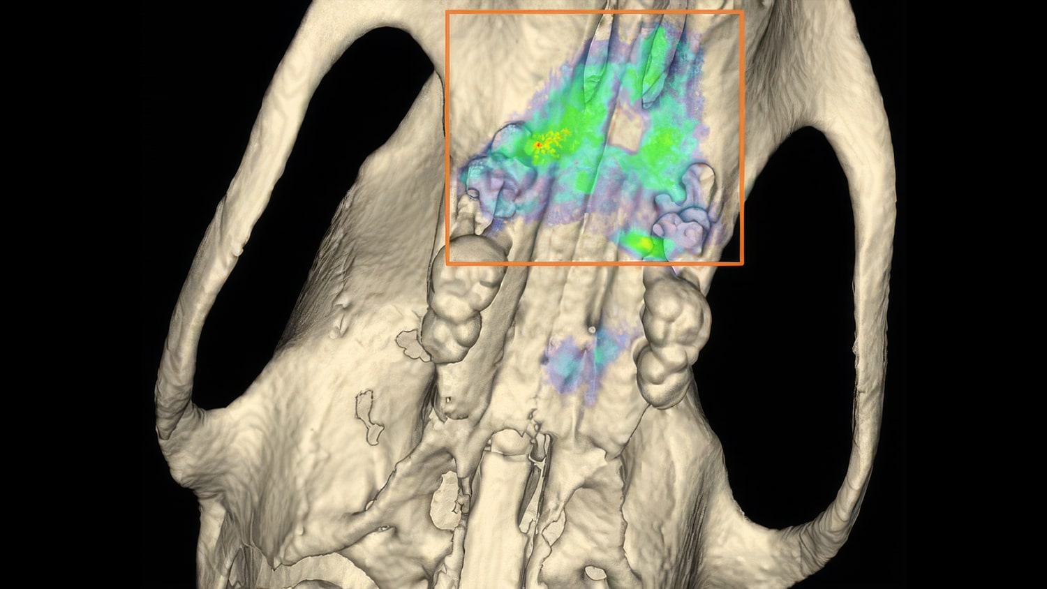
In vivo optical imaging of oral tissue response
Researchers at RWTH Aachen University used integrated optical and anatomical imaging to monitor gingival tissue response following tooth extraction in rodents. The study enabled real-time visualization of tissue changes and comparison of healing dynamics under different experimental conditions.
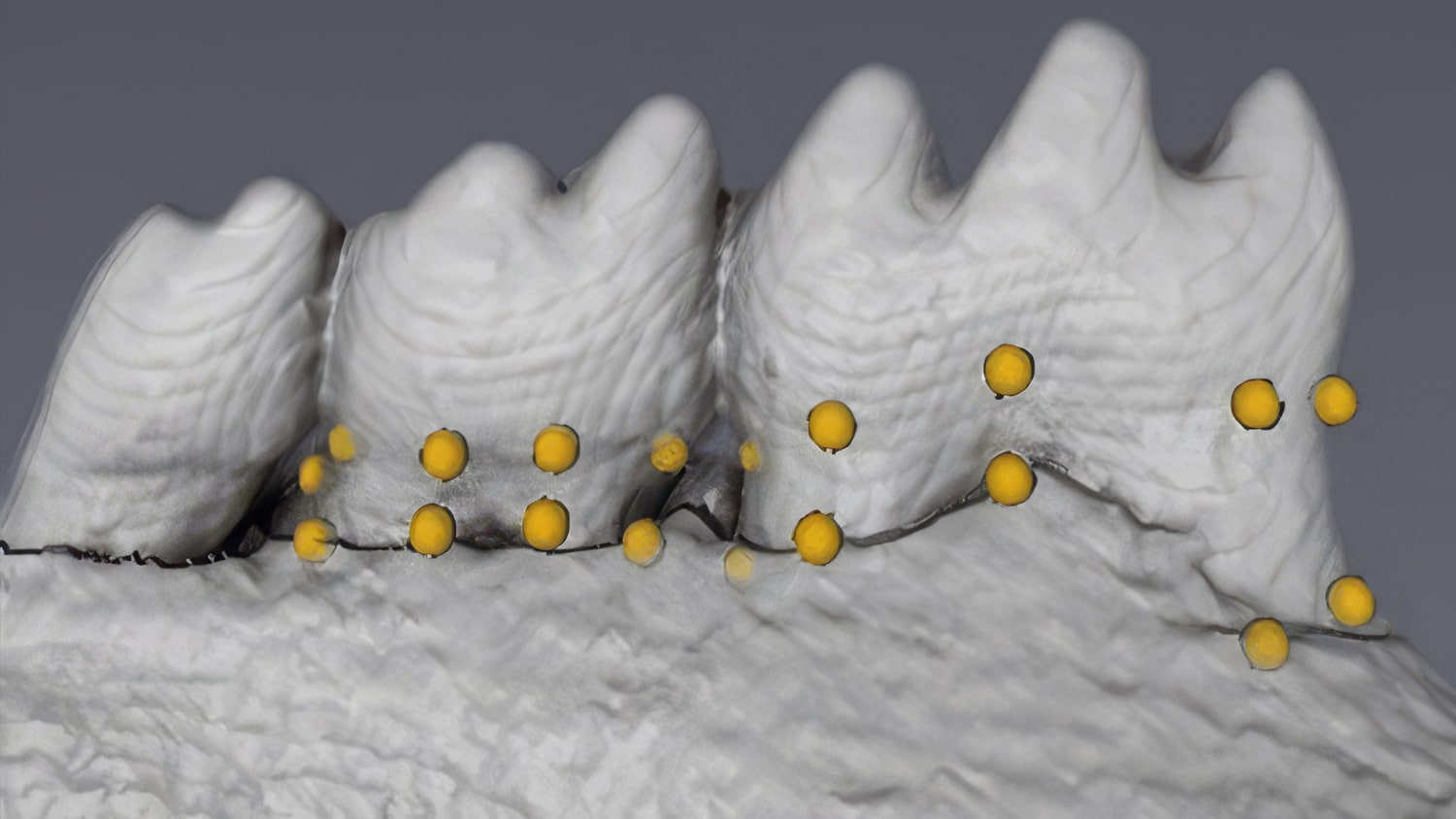
High-resolution 3D imaging of mandibular structure
Researchers at the University of Alberta used ultra-high-resolution CT imaging to generate detailed 3D reconstructions of mandibles, supporting standardized and reproducible assessment of oral structure in preclinical models.