CT imaging for preclinical research
Gain clear, high-resolution anatomical insight fast, so you can quantify structure, track change over time, and strengthen study interpretation.
MILabs’ U-CT systems deliver precise 3D anatomical imaging with exceptional speed and low radiation dose. Used both as a standalone research tool and within multimodal workflows, U-CT supports longitudinal studies and detailed structural analysis across a wide range of research areas and animal models.
Discover our other imaging solutions.
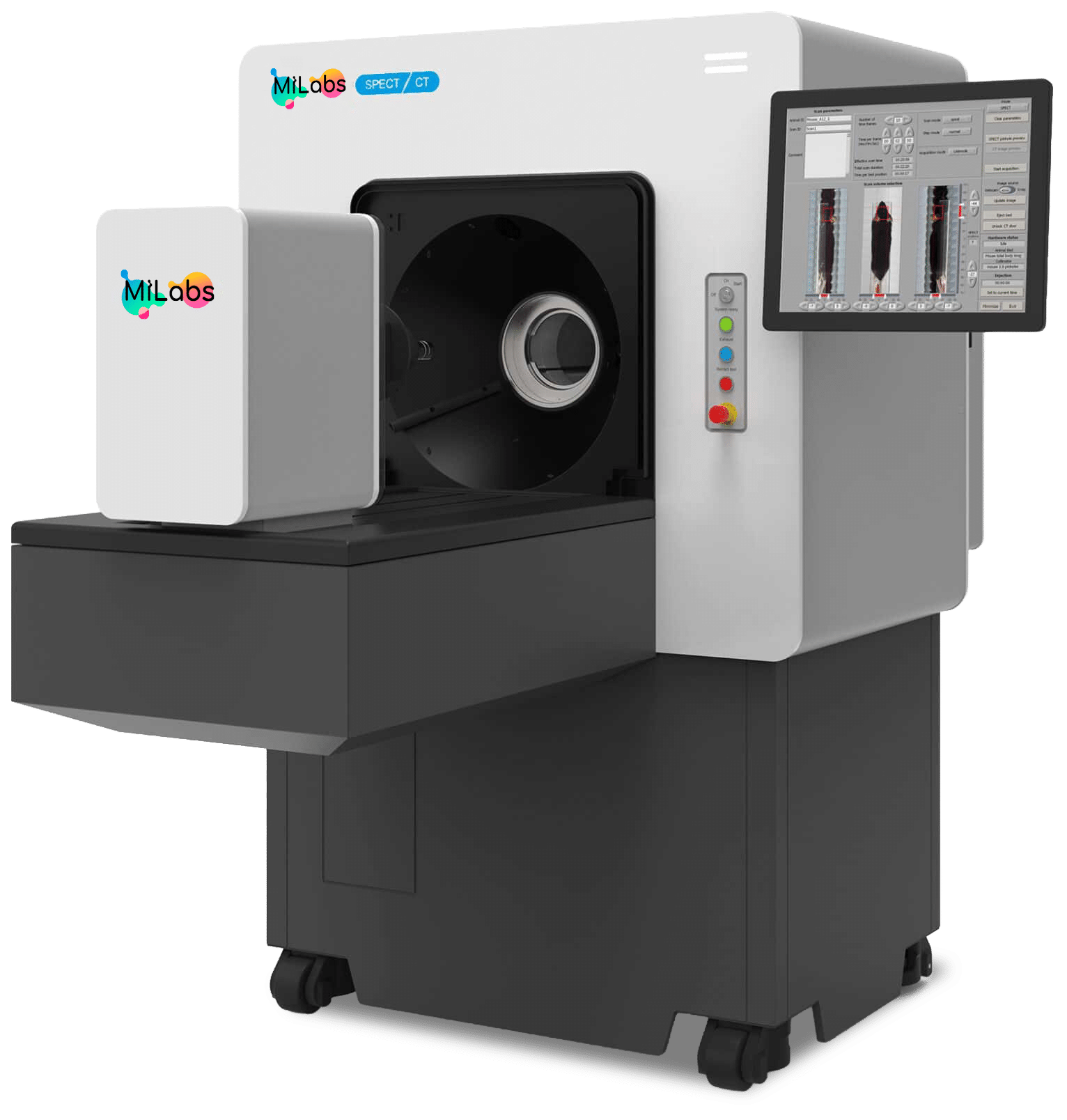
CT scanner from MILabs (U-CT)
Study anatomy as a primary endpoint with speed and precision, so you can quantify structure, track change over time, and generate reliable 3D datasets. With MILabs’ U-CT system, you benefit from:
- EWhole-body mouse imaging in as little as 5 seconds at ultra-low dose (~2 mGy)
- EResolution that scales with your needs, from 30 µm down to 2.4 µm (ex vivo)
- EAdvanced in vivo capabilities (dynamic contrast studies, ultra-fast fluoroscopy, DEXA, virtual endoscopy)
- ESensor-free cardiac and respiratory gating for reliable motion-resolved imaging, up to four mice at a time
- EOptional multi-animal imaging to increase throughput without added complexity

Integrate U-CT with other imaging modalities
Add anatomical context to molecular data and generate aligned, interpretable datasets in a single workflow.
U-CT can be used as a standalone anatomical imaging system or fully integrated with MILabs’ molecular and optical imaging solutions. This flexibility allows researchers to correlate structural and functional information while maintaining image quality and workflow efficiency.
See imaging in action
See how researchers use U-CT to capture ultra-high-resolution bone and anatomical imaging for advanced research applications.
Hear from the experts
Ultra-high resolution in vivo molecular CT system
FAQ
What is the difference between CT and micro-CT?
Micro-CT refers to CT imaging optimized for high-resolution visualization of small structures, making it particularly well suited for preclinical research and materials science.
What is micro-CT used for?
Micro-CT supports detailed 3D reconstruction of anatomical structures for in vivo and ex vivo studies across biology, biomedical research, and materials science.
What are the benefits of U-CT from MILabs?
Researchers benefit from fast scan times, exceptionally low radiation dose, and flexible resolution options, allowing high-quality anatomical imaging that integrates seamlessly into preclinical workflows.