Bone imaging for deeper musculoskeletal insight
See bone structure and biology more clearly, so you can quantify remodeling, vascularization, and metabolic activity with confidence.
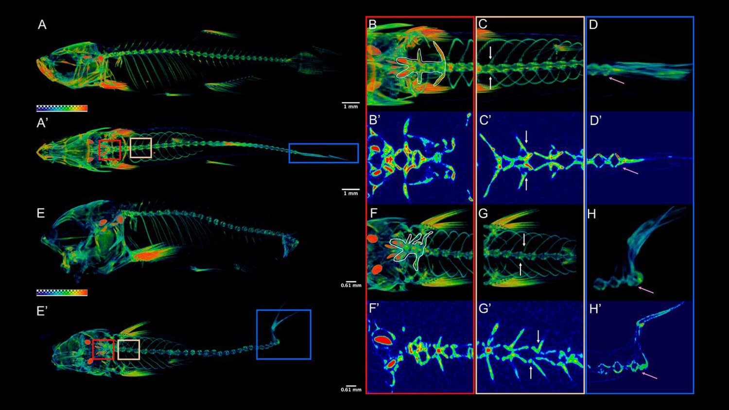
MILabs supports preclinical bone research by enabling researchers to study bone architecture, turnover, and vascular dynamics in vivo and ex vivo. By combining ultra-high resolution anatomical imaging with molecular insight, researchers can generate aligned datasets that support reproducible research across a wide range of musculoskeletal applications.
Discover all other applications.
MILabs equipment for bone imaging applications
Interpret bone biology with greater precision through ultra-high resolution anatomical imaging, with the flexibility to expand into molecular workflows when needed.
MILabs imaging systems provide researchers with scalable tools to study bone structure and biological processes over time. Many teams begin with standalone U-CT for micron-level visualization of bone microarchitecture, remodeling, and vascular structure. When research questions evolve, molecular imaging modalities can be integrated within the same platform to correlate anatomy with tracer-based biological activity. This modular approach allows researchers to reduce variability, strengthen interpretation across longitudinal and interventional study designs, and grow capabilities without replacing their system.
Benefits of MILabs systems for bone research
Ultra-high resolution bone imaging
Resolve fine bone structures and subtle biological changes, so you can study remodeling and microarchitecture with clarity.
MILabs systems support ultra-high resolution imaging that enables detailed visualization of trabecular and cortical bone, supporting studies of bone formation, resorption, and mineralization at small spatial scales.
Dynamic and longitudinal bone studies
Follow bone processes over time, so you can assess biological response and adaptation.
Advanced imaging workflows support dynamic and longitudinal studies of bone remodeling and vascularization. This enables researchers to track spatially correlated changes and evaluate biological responses to interventions with greater confidence.
Integrated molecular and anatomical imaging
Correlate bone structure with biological activity, so you can interpret results within their anatomical context.
By combining molecular imaging with high-resolution CT, researchers can map tracer uptake, metabolic activity, or vascular signals directly onto bone anatomy. This integrated approach supports more comprehensive analysis while reducing study complexity.
Multi-tracer imaging for comprehensive bone biology insight
Compare complementary molecular signals within a single study, so you can assess bone structure, turnover, and metabolism with greater confidence.
MILabs supports multi-tracer bone imaging by enabling aligned SPECT and PET measurements within the same workflow. Using the VECTor platform, researchers can image and compare complementary tracers such as ¹⁸F-NaF and ⁹⁹ᵐTc-HDP, correlating molecular activity with high-resolution bone anatomy. This approach reduces variability between scans, strengthens quantitative interpretation, and supports more robust evaluation of bone remodeling and metabolic processes.
Hear from the experts
Studying osteoarticular biology with molecular imaging
In this webinar, Professor Harrie Weinans (UMC Utrecht) discusses how integrated molecular and anatomical imaging supports research into joint biology and bone remodeling processes.
Imaging musculoskeletal biology with multimodal imaging
Researchers use MILabs imaging platforms to study bone and musculoskeletal biology in vivo and ex vivo, including applications involving bone-implant interactions and soft tissue correlation.