Neurology imaging for clearer understanding of brain systems
See neurological biology more clearly, so you can understand brain systems, quantify molecular signals, and build stronger evidence from in vivo studies.
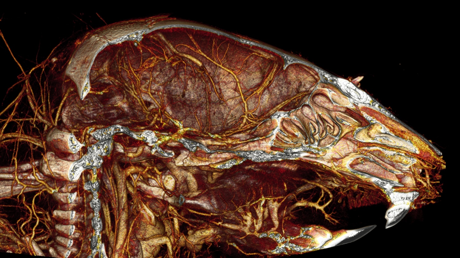
MILabs equipment for neurology applications
Interpret brain biology with greater confidence through high resolution, aligned molecular and anatomical insight in one workflow.
MILabs’ imaging systems provide neurology researchers with an efficient, easy-to-use way to capture high-resolution, quantifiable data from the brain. With flexible molecular and anatomical imaging configurations, researchers can generate richer datasets and support reproducible outcomes across a wide range of study designs.
Benefits of MILabs systems for neurology applications
Ultra-high-resolution capabilities
Resolve small brain structures and faint molecular signals, so you can study neurobiology with greater precision.
MILabs systems support ultra-high-resolution imaging that enables quantitative measurement of receptors and molecular activity in small brain regions, including studies requiring fast kinetics.
Integrated molecular and anatomical imaging
Move faster and compare results with confidence by capturing aligned datasets that reduce study complexity.
By combining molecular and anatomical imaging within a unified workflow, researchers can correlate tracer signals with anatomical structure and acquire multiple datasets in one session.
Multi-tracer imaging for richer neurological insight
See more in a single experiment by comparing complementary molecular signals under identical conditions.
Using the VECTor platform, researchers can perform multi-tracer brain studies that combine SPECT- and PET-based tracers, such as FDG alongside neurotransmitter or receptor-specific tracers. Imaging multiple tracers within the same scan reduces biological variability, improves quantitative comparison, and enables deeper insight into interactions between metabolic activity and specific neurological pathways.
Automated 3D autoradiography
Generate detailed volumetric brain maps without manual sectioning, so you can increase consistency and reduce workflow burden.
EXIRAD-3D automates 3D autoradiography of brain tissue using an efficient scanning workflow. This eliminates common challenges of traditional tissue sectioning and helps teams streamline studies while improving reproducibility and data quality.
Overcoming limitations of MRI
Improve visibility of vascular and structural detail, so you can interpret anatomy and functional biology more reliably.