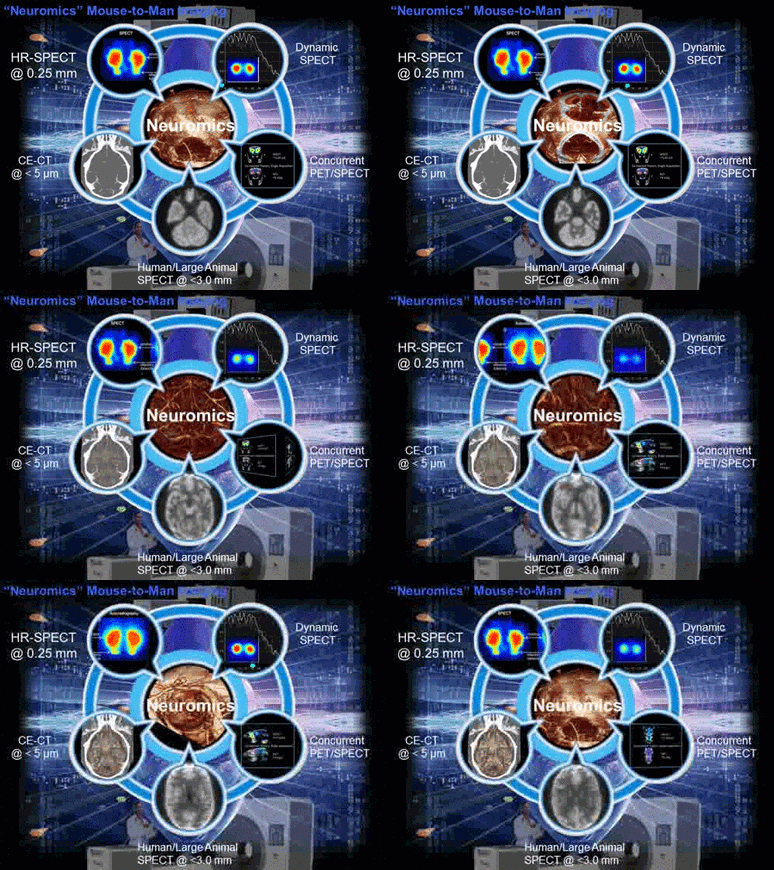
Neuromics: Matters for serious thought…
MIlabs introduces multimodal Neuromics Imaging
Major recent improvements in a number of imaging techniques enable studying the brain in ways that could not be considered previously. Today, with MILabs’ new multimodality neuromics platform, researchers have well-developed and complementary tools for in vivo structural and functional imaging of the rodent brain in action. More specifically, now you can:
| • | Examine the dynamic nature of vascular responses and brain perfusion. |
| • | Study neuroreceptor kinetics at ultra-high resolution with SPECT in species ranging from mice to large non-human primates. |
| • | Gain new insights into brain (dys)functions by simultaneously performing spatially and temporally co-registered PET and SPECT acquisitions. |
| • | Probe neurodegeneration with in vivo bioluminescence and fluorescence imaging. |
| • | Replace labor-intensive and error-prone neural tissue examinations by automated 3D multi-tracer autoradiography. |
| • | Perform SPECT/CT and PET/CT at ultra-high resolutions critical for studying structures that fall well below the threshold of other preclinical hybrid scanners. |
Best of all, you just need one single imaging platform, the MILabs PET/SPECT/OI/CT (VECTor-OI/CT) system, to make this all possible.
 |
| MILabs’ VECTor-OI/CT system makes anatomical, functional and molecular neuromics evaluation possible in a completely non-invasive way. |
Some specific unique applications of the VECTor-OI/CT Neuromics platform include:
High-resolution, low-dose cerebral CT angiography and neurovascular imaging
By using an autonomous gantry on a scalable integrated multimodality platform, MILabs has made drastic improvements in microCT performance. Whether used as stand-alone CT or hybrid molecular CT, the VECtor4OI/CT offers high-resolution scanning efficiency and velocity at ALARA dose levels. These unique 4D CT capabilities make the Neuromics platform suitable for rapid qualitative and quantitative evaluation of mouse cerebral vascular physiology and hemodynamics. Visualizing the 3D architecture of blood vessels with multimodal brain imaging is often crucial since the progression of various neuropathologies ranging from Alzheimer’s disease to brain tumors involves anomalous blood vessels.
Neuroreceptor and neuropsychiatric disorder imaging
For molecular neuroreceptor imaging, MILabs VECtor4OI/CT pushes the limits of what can be achieved in vivo into terrain that was previously only accessible in vitro. Now you can obtain 4D SPECT images with high sensitivity at 0.25 mm resolution, enabling quantitative imaging of receptors in smaller brain areas and with faster kinetics – two capabilities that set MILabs SPECT apart from any other preclinical SPECT imaging system available today. These unique abilities make it possible to image in vivo changes in mouse brain biochemistry such as Aβ deposition, neurotransmitter turnover and metabolism and enable researchers to better understand the pathology mechanisms underlying central nervous system diseases such as Epilepsy, Parkinson’s, NeuroPsych, Alzheimer’s and Stroke.
PET/MRI-counterpart imaging with contrast-enhanced PET/CT and SPECT/CT
By combining the latest in microCT technology with recent advances in contrast agent development and the implementation of analytical software for attenuation-corrected PET/CT and SPECT/CT fusion images, the number of potential applications for contrast-enhanced hybrid nuclear/CT brain imaging has been vastly expanded, often beyond the capabilities of PET/MRI. Indeed, for preclinical research, the use of MRI is often hindered by significantly longer acquisition times to achieve similar levels of resolution than CT, thus reducing throughput. And unlike for brain MRI, X-ray CT imaging techniques are flow independent and therefore issues of non-uniform signal intensity and vessel visibility, caused by variable blood flow, are automatically overcome, thus making VECTor-OI/CT a fast and easy-to-use brain imaging tool for capturing anatomical structure and biologic function data.
Neural tissue analysis with automated 3D multi-tracer autoradiography and Microfil CT
By exploiting the high focusing power of MILabs’ Stationary Multi-Pinhole (SMP) SPECT for smaller ex vivo samples such as the brain, the VECTor-OI/CT Exirad-3D collimator can be used to automate 3D autoradiography applications. Exirad-3D can be used with multiple tracers and eliminates all tissue sectioning–related challenges of brain autoradiography. Moreover, murine brains can be perfused with MICROFIL®(FlowTech Inc., MA), thus enabling the combination of ex vivo functional SPECT mapped of the murine brain vasculature obtained with CT. As such, Exirad-3D SPECT/CT provides a direct link between in vivo and highly detailed ex vivo brain images of the same animal.
In Vivo Optical Imaging in the Study of Neurodegenerative Diseases
The burgeoning choice of optical probes and transgenic animal models are giving in vivo optical brain imaging technology tremendous traction for the assessment of neuropathologies. While conventional in vivo optical imaging is able to provide information on specific reporter location, it is not without a number of practical hurdles. One of the most challenging hurdles is the limited spatial resolution in tissue due to absorption and the difficulty in ascribing depth to the reporter signal as images are commonly acquired as planar and two-dimensional.
Translational nuclear brain imaging with Concurrent PET/SPECT – clinical SPECT redefined
Both PET and SPECT enable the visualization and quantification of specific molecules in the living brain. However, both PET and SPECT are ultimately limited by spatial resolution and the specificity of the radiotracer. Therefore, translating in vivo nuclear imaging findings from laboratory animals to humans remains a considerable challenge. To address these issues, MILabs’ Neuromics platform offers two outstanding capabilities: 1) With Concurrent PET/SPECT acquisitions, researchers can exploit the ultra-high resolution of SPECT for murine brain imaging and switch to PET for translational clinical imaging, by simultaneously acquiring spatially and temporally registered images of the PET and SPECT radiotracers, and 2) A gyro-free G-SPECT imaging platform for large subjects, including humans, will enable isotropic resolutions of <3 mm and dynamic SPECT acquisitions at much reduced radiotracer doses.
