fbpx

← All Categories

Back to all webinars
Bruce Hasegawa Memorial Lecture 2021
This annual lecture honors the legacy of Dr. Bruce Hasegawa. MILabs’ Founder and CEO, Prof. Freek Beekman, presents the 2021 Bruce Hasegawa Memorial Lecture at the University of California, San Francisco. In this video presentation, he talks about the physics, engineering, and application of Fully Integrated Ultra-High Definition Optical Tomography, PET, SPECT & X-ray CT.
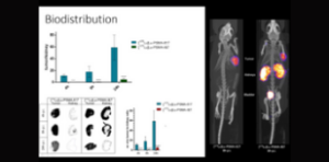
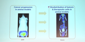
Quantitative hybrid fluorescence and CT to assess drug biodistribution in cancer research
For a detailed biodistribution analysis, PET and SPECT are usually preferred as they are more accurate and quantitative, so the question is, can optical imaging become a complementary tool for pre-clinical biodistribution analysis.
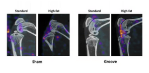
Study osteoarthritis with novel imaging in small animals
The webinar provides an overview of the key role of SPECT/CT imaging on osteoarthritis and rheumatoid arthritis research in small animals.
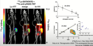
Imaging DNA damage
Each DNA damage repair pathway leads to the recruitment, upregulation, or activation of specific proteins within the nucleus, which, in some cases, can represent attractive targets for molecular imaging.
MILabs VECTor/CT: Applications at the University of British Columbia (UBC)
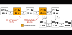
This webinar highlights some of the key imaging applications at the UBC such as theranostic α-emitters imaging (e.g. 225Ac and 211At), nanomedicine, quantitative imaging of novel dual-isotope combinations, such as 111In and 67Ga, investigations of mechanisms of delivery and pharmacokinetics of antibiotics and antimicrobials, investigation of metal chelators and their use as radiopharmaceuticals, studies in immunology and vaccines, and macromolecular conjugates and lipid-based formulations.
Imaging SARS-CoV-2 attacks mitochondria in cardiopulmonary cells in COVID-19 pneumonia
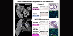
Interested in advancing your drug discovery, pathobiological studies and diagnosis of COVID-19 as well as other progressive pulmonary diseases affecting lung microarchitecture? This webinar provides a comprehensive overview of research in Prof. Stephen Archer’s team, and a good example of their translational approach by incorporating in vivo imaging of mouse models using MILabs’ Micro-CT.
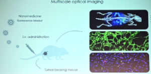
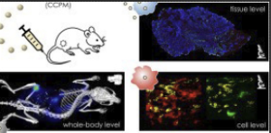
Optical imaging of the whole-body to cellular biodistribution of clinical-stage PEG-b-pHPMA
This webinar describes a multiscale and multimodal optical imaging approach for evaluating core-crosslinked polymeric micelles (CCPM) designed for cancer treatment. The biodistribution and target site accumulation of CCPM were macroscopically and microscopically investigated.
Extensive preclinical evaluation of lutetium-177-labeled PSMA-specific tracers for prostate cancer radionuclide therapy
This talk describes the extensive preclinical evaluation of lutetium-177-labeled PSMA-specific tracers for prostate cancer radionuclide therapy. In vivo imaging was performed on the MILabs VECTorCT imaging platform.
IMplant Infection Therapy (IMIT) ‘RIT for planktonic bacteria and biofilms’
Implant-associated infections such as periprosthetic joint infections are difficult to treat as the bacteria form a biofilm on the prosthetic material. In this webinar, Prof. Van der Wal shows the possibilities of radioimmunotherapy for non-invasive treatment of implant associated infections.
Synergetic Ultra-high Definition PET, SPECT, X-ray and Optical CT
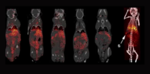
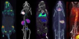
This talk describes the most recent advances of the award-winning MILabs VECTor6 PET, SPECT, X-ray & Optical CT preclinical imaging platform. Several unique applications of the VECTor 6 technology will be presented, performed by many research labs across the globe.
Multifaceted imaging of the musculoskeletal system and more…
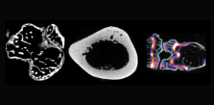
Musculoskeletal imaging plays a major role in disease diagnosis, pathogenesis, and (radio) pharmaceutics development. In this webinar, examples of advanced imaging techniques performed on MILabs’ multimodality PET, SPECT, CT, and OI systems are presented.
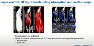
Quantitative Multimodal Fluorescence and Bioluminescence Tomography with the MILabs OI-CT System
Hybrid fluorescence tomography and x-ray computed tomography (FLT-CT) provided by the MILabs OI-CT system allows non-invasive longitudinal volumetric assessment of fluorescent probes in mice in vivo. This talk demonstrates that FLT-CT can be used to quantitatively determine the biodistribution NIRF-labeled probes.
MILabs U-SPECT/CT imaging in immuno-oncology
This webinar describes the key applications of nuclear imaging in immuno-oncology. Currently, numerous immuno-oncological drugs are being developed to treat cancer patients. However, many questions remain unanswered, such as how, when, and for whom this treatment is most effective.
Airway Remodeling in Ferrets with Cigarette Smoke Induced COPD using µCT Imaging
Ferrets are a recently established animal model uniquely exhibiting similar clinical and pathological characteristics of COPD as humans, including chronic bronchitis but are also instrumental for COVID-19 research. This talk describes a µCT method for evaluating structural changes to the airways in ferrets and how the MILabs µCT can be used as a significant translational platform to measure dynamic airway morphological changes.
MILabs VECTor/CT: Applications at the University of Saskatchewan
Radioimmunoimaging and radioimmunotherapy applications are supported by the MILabs VECTorCT platform at the University of Saskatchewan.
MILabs USPECT4CT: applications in a preclinical study of 212Pb alpha radioimmunotherapy in multiple myeloma
This webinar highlights the potency of 212Pb-alpha radioimmunotherapy in Multiple Myeloma treatment. The results of this study showed the efficacy of 212Pb-daratumumab in xenografted mice with significant tumor regression and increased survival.
Dynamic SPECT/CT imaging of glymphatic drug delivery and clearance in small animals
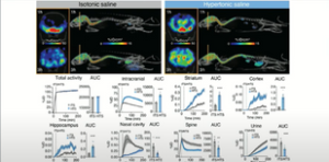
Marko Rosenholm presents some of the findings and results regarding dynamic SPECT/CT imaging of a glymphatic system they have acquired with the MILabs VECTor/CT system.
Dual Isotope Imaging Using the MILabs VECTorCT: Applications in Oncology
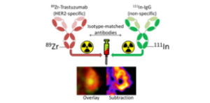
This webinar covers some recent groundbreaking studies conducted by the Cornelissen group in Oxford. Several recent papers will be presented including ‘Early detection in a mouse model of pancreatic cancer by imaging DNA damage response signaling’, ‘Dual-isotope antibody imaging allows in vivo immunohistochemistry using radiolabelled antibodies in tumours’, and ‘Imaging DNA Damage Repair in vivo Following 177Lu-DOTATATE Therapy’.
Multiscale optical imaging for unraveling the in vivo fate of nanomedicine
The Institute for Experimental Molecular Imaging’s (RWTH Aachen) research team uses MILabs Optical imaging system, intravital microscopy, and immunohistochemistry to investigate cancer nanomedicine delivery and uptake in vivo.
Multimodal in vivo imaging of CAR T cell therapies using MILab’s OI-CT system
This webinar describes Miltenyi Biotech’s approach establishing multimodal optical imaging of CAR T cell therapies, using the MILabs optical imaging micro CT system.
Scroll to Top