May 12, 2020, Southampton, UK – Utrecht, the Netherlands
The Faculty of Medicine, University of Southampton have been awarded a large equipment grant from the Biotechnology and Biological Sciences Research Council (BBSRC) under a consortium of investigators led by Professor Richard Oreffo entitled: Correlative In Vivo Fluorescence and Micro-Computed Tomographic Imaging of Tissue Structure and Function.
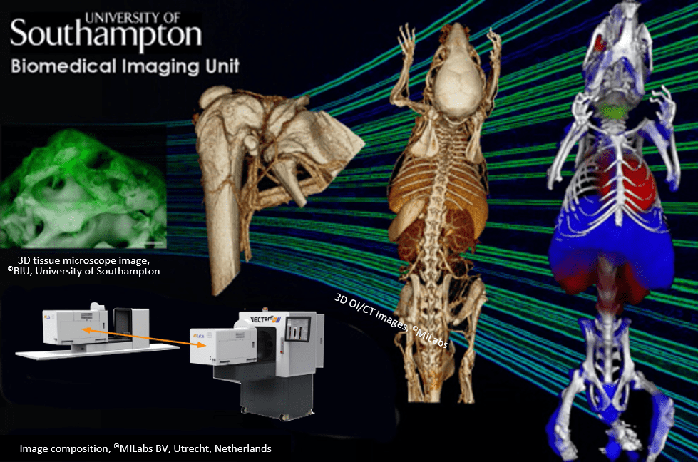
The Biomedical Imaging Unit (BIU) of the University of Southampton has acquired MILabs market-leading Optical-CT imaging system, an integrated, non-invasive, non-destructive imaging system to track the spatial and temporal distribution of tissue structures and physiologically relevant labels in tissue-engineered constructs, and tissue samples in live animals. This unique system features high-resolution CT combined with 2D, 3D and 4D optical imaging to enabling correlative in vivo imaging applications to understand tissue development and formation in its native 3D context.
According to Professor Oreffo, “Correlative in vivo imaging has the potential to transform human health over the next 10-20 years. Our ability to label and follow specific cells and molecules within the body and to create 3D image stacks of tissues of interest with high-resolution micro-computed tomography offers a step change in our research approach to understanding basic physiology and for regenerative medicine approaches.”
According to Dr. Anton Page, Head of the Biomedical Imaging Unit (BIU): “The MILabs system is a multispecies preclinical imaging system, offering longitudinal in vivo imaging capabilities, in addition to ex vivo sample scanning. It is important to note that this system allows not only imaging of hard tissues such as bone but also soft tissues, including lung, liver, adipose tissue, vascular systems, or cancerous tissue. This equipment will compliment 3D imaging capability of the BIU, which already includes light sheet microscopy, confocal, x-ray tomography, serial block face scanning electron microscopy and electron tomography and will allow us to do correlative imaging across the length scales. As a multi-user facility we will be able to provide integrated, non-invasive, non-destructive imaging to track the spatial and temporal distribution of tissue structures and physiologically relevant labels in live animals applicable to a number of research projects across the University”.
Prof. Frederik Beekman, CEO of MILabs adds: “We believe that our Optical imaging system integrated with ultra-high-resolution CT provides a unique tool for Correlative In Vivo Imaging studies. We are excited that this system with CT-guided 3D/4D bioluminescence and fluorescence meets a key unmet challenge at the University of Southampton to track tissue architecture and function non-destructively in living animals.”
About Biomedical Imaging Unit, Southampton:
The Biomedical Imaging Unit is a joint University Hospitals Southampton NHS Foundation Trust (UHS)/University of Southampton (UoS) facility for high quality/high resolution diagnostic and research imaging. Accredited under UKAS to ISO 15189, this core facility carries out research work for UoS, other universities and research institutes, industry, and hospitals. As a multi-user facility supported by ten highly experienced and expert imaging staff, the Biomedical Imaging Unit can provide a complete supporting workflow for internal and external users in all aspects of imaging from experimental design, proof of concept tests, user training, sample preparation, imaging, interpretation, processing, and analysis of specimens, data management and archiving. For more information: https://www.southampton.ac.uk/biu/index.page
About MILabs, B.V.
This fast-growing Dutch company has a history of providing a continuum of innovations to expand the applications of preclinical molecular imaging. With its latest adaptive platform, MILabs has succeeded at commercializing a scalable imaging platform, able to accommodate exclusive Optical tomography and ultra-high-resolution CT imaging as well as high-definition nuclear PET and SPECT techniques. The company has built a strong brand based on its mission of “Making Molecular Imaging Clear” complemented by multiparametric imaging and unique concurrent within subject analysis of multiple probes. it provides efficient translational systems for both diagnostic and therapy applications while minimizing the use of animal models. For more information, visit: www.milabs.com or contact MILabs at [email protected]
