|
Why Stationary SPECT outclasses PET for in vivo animal imaging
|
|
Positron Emission Tomography (PET) and Single Photon Emission Computed Tomography (SPECT) offer the sensitivity and quantitative accuracy required for molecular imaging with extremely low concentration of radiolabeled molecules. Although human clinical PET outperforms current clinical SPECT, the recent advent of full angular preclinical Stationary SPECT (U-SPECT) outclasses PET image quality for many small animal imaging applications. The following examples demonstrate these unique capabilities of current U-SPECT.
|
|
|
1. Unprecedented Power in Musculoskeletal Mouse Imaging
|
|
|
With the ability to image molecules in small bone structures at 140 micon ex vivo and 250 micron in vivo, U-SPECT brings a high level of sophistication to molecular detection of the underlying causes of musculoskeletal diseases; an advancement that appeared futuristic just a couple of years ago. Indeed, U-SPECT enables quantifying molecules in volumes that are 10 to 100 times smaller than with PET.
|
|
|
2. Enabling the Future of Cardiac Research in Mice
|
|
|
MILabs’ dual-gated U-SPECT for cardiac research enables visualizing and quantifying molecular signals from the murine heart invisible with any other molecular imaging technology, including PET. U-SPECT enables imaging, tracking, and quantifying molecular biomarkers in sub-half-mm sized structures of a living heart, such as the atrial wall and papillary muscle, allowing every cardiac imaging test available in humans also in mice. In addition it opens many new horizons in cardiology research.
|
|
|
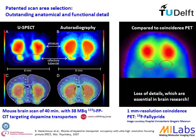
3. Large-scale Functional Imaging of Small Brains
|
|
|
Before the development of MILabs’ U-SPECT, the limited spatiotemporal resolution of nuclear imagers forced researchers to use rats to differentiate small brain structures. U-SPECT now enables to unlock signals in small mouse brain structures associated with e.g. Parkinson’s, Alzheimer’s, Lewi Body dementia, and addiction. Examples comprise quantification of dopamine transporter dynamics in tiny structures such as the olfactory tubercle in mice.
|
|
|
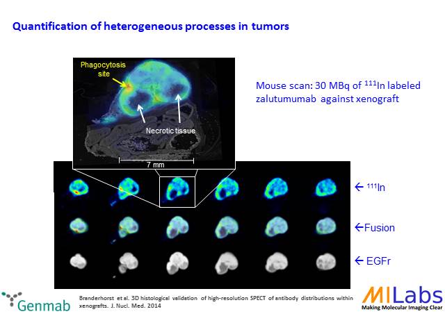
4. Elucidating Treatment Efficacy in Tumor Models
|
|
|
Targeted drug delivery to tumors remains a pharmacological challenge. Dosage of e.g. antibodies in different parts of a tumor is highly variable. U-SPECT enables to quantitatively assess distribution of pharmaceuticals and biomarkers to determine efficiency of e.g. immunotherapy and oncologic virotherapy.
|
|
|
5. Whole-body SPECT Pharmacokinetic Modelling
|
|
|
Dynamic studies – once the privileged domain of PET – can now be performed with U-SPECT by acquiring tomographic images without detector rotation. This enables ultra-fast organ kinetic studies with sub-second time frames as well as whole-body dynamic acquisitions with <10 sec time resolution. Fast and precise, without gantry wear-and-tear, and detector positioning inaccuracies is the only way to guarantee top class, and reproducible longitudinal studies.
|
|
6. Theranostic Imaging at Sub-mm Resolution
|
The ability to use MILabs’ proprietary M7TM ultra-high energy pinhole collimation extends the application to image-guided radionuclide therapy. Virtually all radiotherapy isotopes including Auger e-, alpha-, and beta emitters such as 131I, 188Re, 213Bi, 209At, and 225Ac can now be imaged at sub-mm resolution.
|
|
Unlike any other technology, stationary SPECT linearly scales with the volume of the object (down to zebra-fish with U-SPECT up to large animals with G-SPECT*), so one can image tiny animals with the same visual acuity as humans – without information loss from bench to bedside.
Also, one can use U-SPECT and PET in the same animal simultaneously. Just by upgrading MILabs’ U-SPECT to VECTor with the Adaptive PET Modality. Before converting your study to clinical PET, you can image with both SPECT and PET, and do a one-on-one translation with both spatially and temporally perfectly overlaid PET & SPECT.
|
|
If you still need more proof: we have more than 100. Today MILabs’ U-SPECT and VECTor have already played an instrumental role in several hundreds of published preclinical studies, demonstrating superior performance for a wide range of small animal imaging applications.
* G-SPECT for Human Clinical Scanning depends on final local registrations, including FDA and CE clearance.
|
|
|
EMIM, March 19 – 22 2019, Glasgow
AACR, March 30 – April 3 2019, Atlanta
NukMed, April 3 – 6 2019, Bremen
ISRS, May 26 – 31 2019, Beijing
SNMMI, June 22 – 25 3 2019, Anaheim
EANM, October 12-16, Barcelona
|
|
|