Small Animal Brain SPECT-PET
Unique capabilities of MILabs’ U-SPECT and VECTor systems include the quantification of fast dynamics of both SPECT and PET tracers, due to the absence of rotating detectors. VECTor’s ability to acquire simultaneously sub-mm resolution images of multiple SPECT and PET tracers is a unique benefit for researchers and simple to carry out.
Selected publications:
| ► | High-resolution quantitative PET with minimal partial volume effects is essential for brain imaging. In a recent paper by Walker and colleagues (JNM 2014) the importance of high-resolution and high contrast brain imaging using clustered-pinholes was shown and compared with traditional coincidence PET. | |||||||||||||
|
|
|||||||||||||
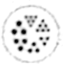
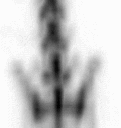
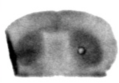
| Images of Jaszczak phantom with rod diameters of 0.85, 0.95, 1.10, 1.30, 1.50, and 1.70 mm. (A) Results for VECTor, (B) Results for coincidence PET using 3DMAP (β=0), for equal duration frames, (C) 18F-NaF bone imaging in pelvis and lower spine of mice. with VECTor and (D) coincidence PET using 3DMap (β=0), (E) 18F-FDG image acquired on VECTor and F. corresponding ex vivo autoradiography of mouse brain [1]. | ||||||||||||||


| ► | Ultra-fast pharmacokinetic SPECT is essential in the evaluation of a selection of serotoninergic tracers. Tsartsalis and colleagues (Molecular Imaging 2014) show exciting results with U-SPECT/CT. | |
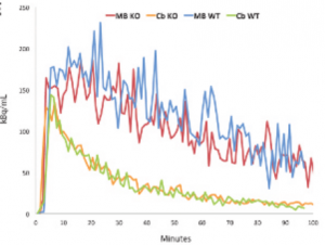 |
||
| Regional time-activity curves measured by SPECT after tracer injection in the rat brain [2]. | ||
| ► | High resolution subtraction SPECT imaging as is clinical in use for determining epileptogenic zones in the brain can also be used accurately locate the spatial extent and intensity of perfusion change after brain stimulation. | |
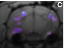 |
||
| Subtraction analysis of HMPAO-Tc99m SPECT tracer showing perfusion changes after brain stimulation [3]. | ||
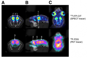
| ► | High resolution simultaneous SPECT and PET image with VECTor provides unique complementary information about tissue function. These combined unique capabilities provide new perspectives for life science research. | |
 |
||
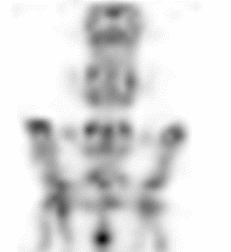
| Simultaneously acquired SPECT/PET mouse brain images (color) overlayed on CT (gray). Coronal (A), sagittal (B), and horizontal (C) slices at identical levels are shown. Mouse was injected with 30 MBq of 123I-FP-CIT and 40 MBq of 18F-FDG and imaged for 60 min starting 105 min after injection. Uptake of 123I- FP-CIT (SPECT, top) in small brain structures such as striatum (1), olfactory tubercle (2), and Harderian glands (3) can be resolved. 18F- FDG (PET scan, bottom) is seen in multiple structures, including olfactory bulbs (4), cerebral cortex (5), and thalamic and midbrain areas (6) [4]. | ||
| For example VECTor is an enabling technology for use in a variety of research areas in which a direct correlation of multiple biologic functions will lead to enhanced insights. Two tremendously large and important classes of tracers (PET and SPECT) can now be combined: | ||
| • | VECTor can play also a significant role in simultaneous and possibly dynamic imaging of different brain processes, such as metabolic activity in defined brain regions together with the functioning of the neuro- transmitter system (—or, alternatively, in monitoring various aspects of the neurotransmitter system simultaneously. Specifically, many research groups are currently investigating the effect of an intervention (e.g., amphetamine or cocaine administration) on metabolic brain activity (monitored with 18F-FDG) or on the neurotransmitter system (imaged with SPECT tracers such as IBZM, FP-CIT, or TRODAT). Because the biologic system does not react in the same way to repeated interventions (an effect called sensitization), these 2 effects can be directly correlated only through simultaneous SPECT/PET. Furthermore, in research related to Parkinson disease, the possibility of using simultaneous SPECT/ PET to image multiple sites or processes of interest—thereby enabling a direct investigation of interactions between different processes—has already attracted interest. | |
| • | VECTor can also be used in tracer research to exactly compare a SPECT and a PET tracer that target the same biologic function. In such a simultaneous SPECT/PET scan, the targeting properties of a molecule labeled with a heavy metal—such as is commonly used for SPECT tracers—could be compared directly with the behavior of an identical molecule labeled with a small positron- emitting radioactive atom. Such a comparison, at the same time and at exactly the same position in a single animal, rules out the effects of varying physiology and spatiotemporal misregistration—effects that today severely complicate tracer comparisons. This, in turn, can aid in the modification of a PET tracer into a SPECT tracer. The modification of PET tracers into SPECT tracers is of practical interest because of the wider availability of SPECT and may also be necessary when a process needs to be monitored for a long time—something prohibited by the relatively short lifetimes of most PET labels. | |
References
| 1. | Performance Assessment of a Preclinical PET Scanner with Pinhole Collimation by Comparison to a Coincidence-Based Small-Animal PET Scanner, Walker et al. Journal of Nuclear Medicine, p1368-1374, 2014 |
| 2. | Small-animal single-photon emission computed tomographic imaging of the brain serotoninergic systems in wild-type and mdr1a knockout rats. Dumas et al. Molecular Imaging, p1-12, 2014 |
| 3. | Hippocampal deep brain stimulation induces decreased rCBF in the hippocampal formation of the rat, Wyckhuys et al. NeuroImage p55-61, 2010 |
| 4. | VECTor: A Preclinical Imaging System for Simultaneous Submillimeter SPECT and PET, Goorden et al. Journal of Nuclear Medicine, p306-312, 2012 |
